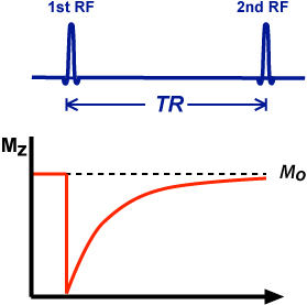
Posts

Showing posts from 2018

Radiation

Image
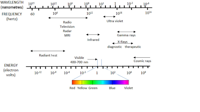
Radiation is the emission or transmission of energy in the form of waves or particles through space or through a material medium. Electromagnetic radiation Electromagnetic waves can, like all waves, be characterized by their amplitude, wavelength ( λ ), frequency ( ν ) and speed. The amplitude is the intensity of the wave. The wavelength is the distance between identical points on adjacent cycles. The frequency is the number of complete wave oscillations per unit time. The speed of the wave is equal to the product of the frequency and the wavelength, and its magnitude depends upon the nature of the material through which the wave travels and the frequency of the radiation. In a vacuum, however, the speed for all electromagnetic waves is a constant, usually denoted by c , and  in which case: c = λν For X rays, wavelength is usually expressed in nanometres (nm) (1 nm =  10 -9  m) and frequency is expressed in Hertz (Hz) (1 Hz = 1 cycle/s = 1 s–1). Whe

Relaxation

Image
Relaxation MRI image contrast is influenced by several characteristics of tissues and other materials including: T1, T2 and T2* relaxation as well as spin density, susceptibility effects and flow effects. This page and the following detailed pages on relaxation parameters will introduce properties and effects on imaging contrast, time and signal-to-noise ratio (SNR). Relaxation  is the process in which spins release the energy received from a radiofrequency pulse. T1 and T2 relaxation rates affect the SNR in an image. Improvement in the SNR is seen when the TR is increased significantly to about 3-5 T1 times. Changing the TR time will also affect the T1 weighting of the image and the acquisition time. T1 weighting occurs in a short TR spin echo sequence because of incomplete recovery of longitudinal magnetization. The effect of TR on SNR can be shown graphically by a T1 relaxation curve as illustrated in the exponential growth curve in figure 1. As the TR is increased to

Electromagnetic Induction

Image
Electromagnetic induction Electromagnetic induction  is the induction of electric current via changing magnetic fields.  Magnetic fields  are generated by moving charges (equivalent to electrical current). Ampere’s law or Fleming’s right hand rule determines the magnitude and direction (i.e. clockwise or anti-clockwise) of the magnetic field with respect to the direction of the flow of current. That is, if you point your right thumb in the direction of the current, the magnetic field will follow the curve of your fingers. Changing magnetic fields can induce an electric current. When a magnet is moved in and out of a closed circuit, an oscillating current is produced which ceases the moment the magnet stops moving. This is explained by Faraday’s law of induction. The change in magnetic flux through a closed circuit induces an electromotive force (EMF) in the circuit. The EMF drives a current in the circuit. The laws of electromagnetic induction state that the induced EMF is:

Larmor Frequency

Larmor Frequency The  Larmor frequency  and equation are named after the Irish physicist and mathematician  Joseph Larmor  (1857-1942). Protons and neutrons pair up in nuclei causing the cancellation of their individual angular momentum. All nuclei also have a spin; those with an odd number of protons and/or neutrons will have a property called a magnetic (dipole) moment. Magnetic moment is characterized by its alignment with an external magnetic field analogous to a small bar magnet. These particular nuclei are also called dipoles because they have two poles like the north and south pole of a bar magnet. H-1 and P-31 are examples of nuclei with an unpaired proton. N-14 is an example of a nucleus with both an unpaired proton and neutron. In addition to alignment of nuclei with a magnetic moment, application of an external magnetic field will produce a secondary spin or wobble ( precession ) of nuclei around the main or static magnetic field. The precessional path around the m

Repetition time

Image
Repetition time    The repetition time (TR) is the time from the application of an excitation pulse to the application of the next pulse. It determines how much longitudinal magnetization recovers between each pulse. It is measured in milliseconds.

Flip Angle

Image
FLIP ANGLE    The flip angle is an MRI phenomenon by which the axis of the hydrogen proton shifts from its longitudinal plane (static magnetic field B0)Z axis to its transverse plane XY axis by excitation with the help of radiofrequency (RF) pulses .  A RF pulse is sent in at the precise Larmor frequency in relation to the gyromagnetic ratio and magnetic field strength.

Echo Time

Image
ECHO TIME The echo time (TE) refers to the time between the application of the radiofrequency excitation pulse and the peak of the signal induced in the coil. It is measured in milliseconds. The amount of T2 relaxation is controlled by the TE.

MRI Physics

Image
THE BASIC PROCESS The way MR images are generated is complicated and is much harder to understand than plain radiography, CT and ultrasound. It has strong underpinnings in physics which must be understood before any real sense of 'how it works' is gained. What follows is a very abbreviated, 'broad strokes' description of the process. Essentially, the process can be broken down into four parts: 1. Preparation 2. Excitation 3. Spatial encoding 4. Signal acquisition Preparation The patient is placed in a static magnetic field produced by the magnet of the MR scanner. In living tissues there are a lot of hydrogen atoms included in water molecules or in many different other molecules. The proton, the nucleus of hydrogen, possesses an intrinsic magnetisation called spin. The spin magnetization vector precesses (rotates) around the magnetic field at a frequency called the Larmor frequency, which is proportional to the magnetic field intensity. The

Why use MRI

Advantages : Advantages of MRI include:  ability to image without the use of ionising x-rays, in contradistinction to CT scanning images may be acquired in multiple planes (axial, sagittal, coronal, or oblique) without repositioning the patient. CT images have only relatively recently been able to be reconstructed in multiple planes with the same spatial resolution MRI images demonstrate superior soft tissue contrast as compared to CT scans and plain radiographs making it the ideal examination of the brain, spine, joints, and other soft tissue body parts some angiographic images can be obtained without the use of contrast material, unlike CT or conventional angiography advanced techniques such as diffusion, spectroscopy and perfusion allow for precise tissue characterisation rather than merely 'macroscopic' imaging functional MRI allows visualisation of both active parts of the brain during certain activities and understanding of the underlying networks. Disa

MRI

Image
Magnetic resonance imaging (MRI) is a medical imaging technique used in radiology to form pictures of the anatomy and the physiological processes of the body in both health and disease. MRI scanners use strong magnetic fields, magnetic field gradients, and radio waves to generate images of the organs in the body. MRI does not involve X-rays or the use of ionizing radiation, which distinguishes it from CT or CAT scans and PET scans. Magnetic resonance imaging is a medical application of nuclear magnetic resonance (NMR). NMR can also be used for imaging in other NMR applications such as NMR spectroscopy. MRI (an abbreviation of magnetic resonance imaging) is an imaging modality that uses non-ionising radiation to create useful diagnostic images. MRI was initially called nuclear magnetic resonance (NMR) imaging after its early use for chemical analysis. The initial " nuclear " part was dropped about 25 years ago because of fears that people would think there was some

Skeleton

Image
Skeleton includes bones and cartilages. It forms the main supporting framework of the body, and is primarily designed for a more effective production of movements by the attached muscles. BONES Synonyms 1. Os (L) 2.Osteon (G) Compare with the terms, osteology, ossification, osteomyelitis, osteomalacia, osteoma, etc Definition     Bone  is  one-third  connective  tissue.  It  is  impregnated  with  calcium salts  which  constitute  two-thirds  part.  The  inorganic  calcium  salts (mainly  calcium  phosphate,  partly  calcium  carbonate,  and  traces  of other  salts)  make  it  hard  and  rigid,  which  can  afford  resistance  to compressive  forces  of  weight-bearing  and  impact  forces  of  jumping. The  organic  connective  tissue  (collagen  fibres)  makes  it  tough  and resilient  (flexible),  which  can  afford  resistance  to  tensile  forces.  In strength,  bone  is  comparable  to  iron  and  steel.    Despite  its  hardness  and  high  calcium  content  t

Introduction to the Human Body

Image
Human anatomy is the study of the shape and form of the human body. The human body has four limbs(two arms and two legs), a head and a neck which connect to the torso. The body's shape is determined by a strong skeleton made of bone and cartilage, surrounded by fat, muscle, connective tissue, organs, and other structures. The spine at the back of the skeleton contains the flexible vertebral column which surrounds the spinal cord, which is a collection of nerve fibers  connecting the brain to the rest of the body. Nerves connect the spinal cord and brain to the rest of the body. All major bones, muscles, and nerves in the body are named, with the exception of anatomical variations such as sesamoid bones and accessory muscles. Blood vessels carry blood throughout the body, which moves because of the beating of the heart. Venules and veins collect blood low in oxygen from tissues throughout the body. These collect in progressively larger veins until they reach the body&#
Welcome to Radiological Assistance page. Here you can find some interesting fact about Radiology.